Protein Structure
Follow the same amino acids from sequence to helices, sheets and an experimental fold.
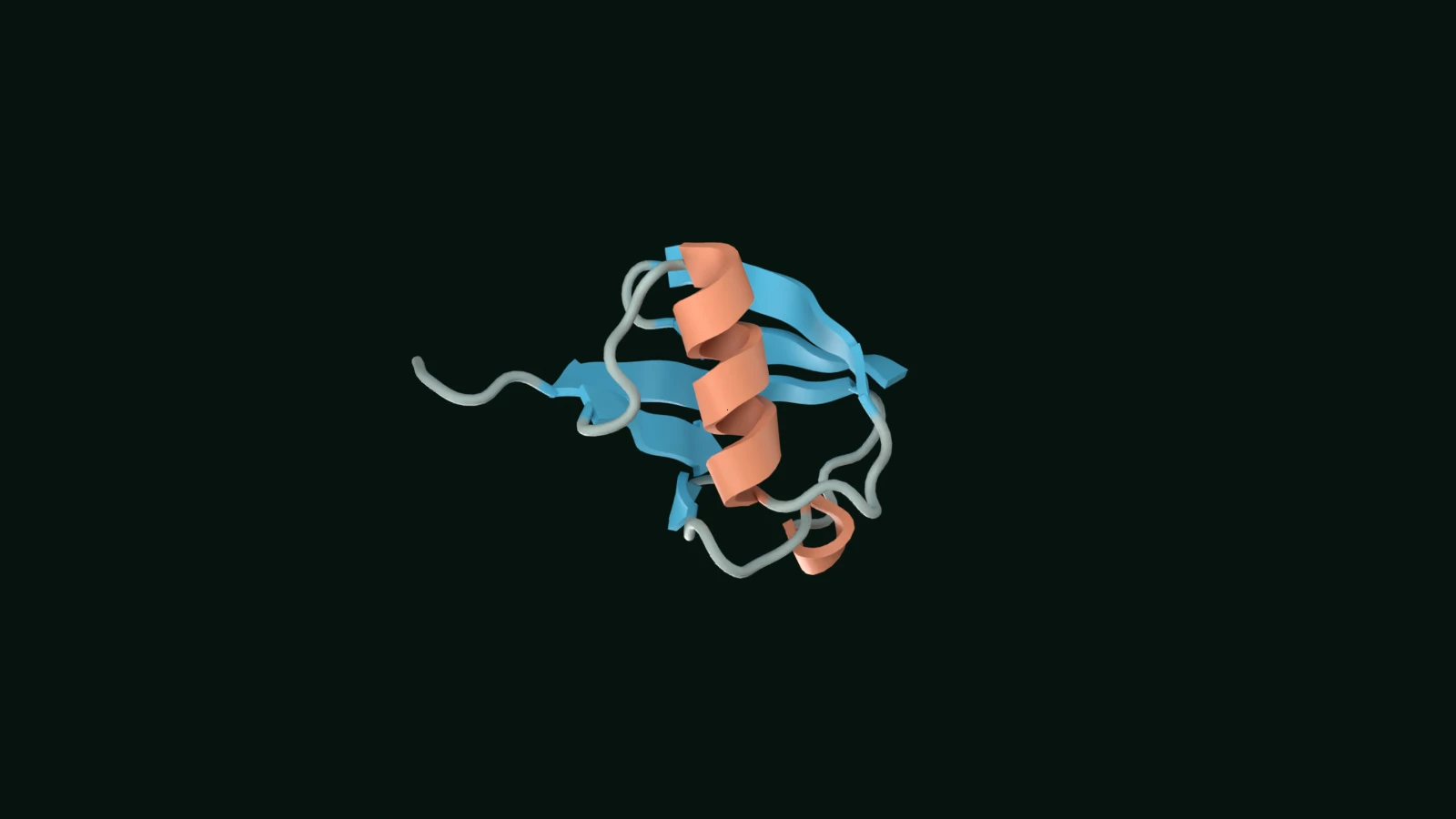
Loading the experiment…
Quick reference
One chain, several structural levels
Sequence records the order of amino acids. Local backbone arrangements form helices and sheets, while the full chain packs into a three-dimensional fold. A view of the sequence and a view of the fold describe the same molecule.
A small experimental example
Human ubiquitin in PDB 1UBQ has 76 residues, a main alpha helix, a short 3₁₀ helix and a mixed five-stranded beta sheet. The experiment highlights the deposited assignments so you can compare sequence positions with their arrangement in space.
Read evidence with its meaning
This is an X-ray structure refined at 1.80 Å resolution. Its B factors describe atomic displacement in that crystallographic model; they are not prediction-confidence percentages. The final four residues have greater disorder. A single structure also does not show every conformation available to a protein.
Check your understanding Optional
Two beta strands are far apart in sequence but adjacent in the structure. What does that show?
At the bench
Use Protein to inspect experimental structures, predicted models, sequence annotations and source evidence for proteins relevant to your work.
Open ProteinSources and model notes
Structure-based views of resolved experimental coordinates. Cutaways, category colours, camera choices and guided highlights are authored for teaching. Coordinates remain fixed; highlights are not experimental trajectories or simulation outputs.
Experimental coordinates, 76-residue sequence and primary structural publication. Feature ranges come from the deposited mmCIF assignments.
How experimental evidence supports structural models.