Binding and Inhibition
Explore a ligand pocket, then change affinity and concentration in an equilibrium binding model.
Loading the experiment…
Quick reference
Affinity and occupancy
At equilibrium, a single-site binding model gives half occupancy when the free ligand concentration equals its dissociation constant, Kd. A lower Kd means higher affinity under the stated conditions. Occupancy also depends on how much free ligand is available.
Competition changes the balance
A second ligand competing for the same site reduces occupancy by the first. The concentration needed to halve that occupancy depends on the first ligand's concentration and affinity as well as the competitor's Ki. The sliders prescribe free concentrations and omit depletion.
A structure is a different kind of evidence
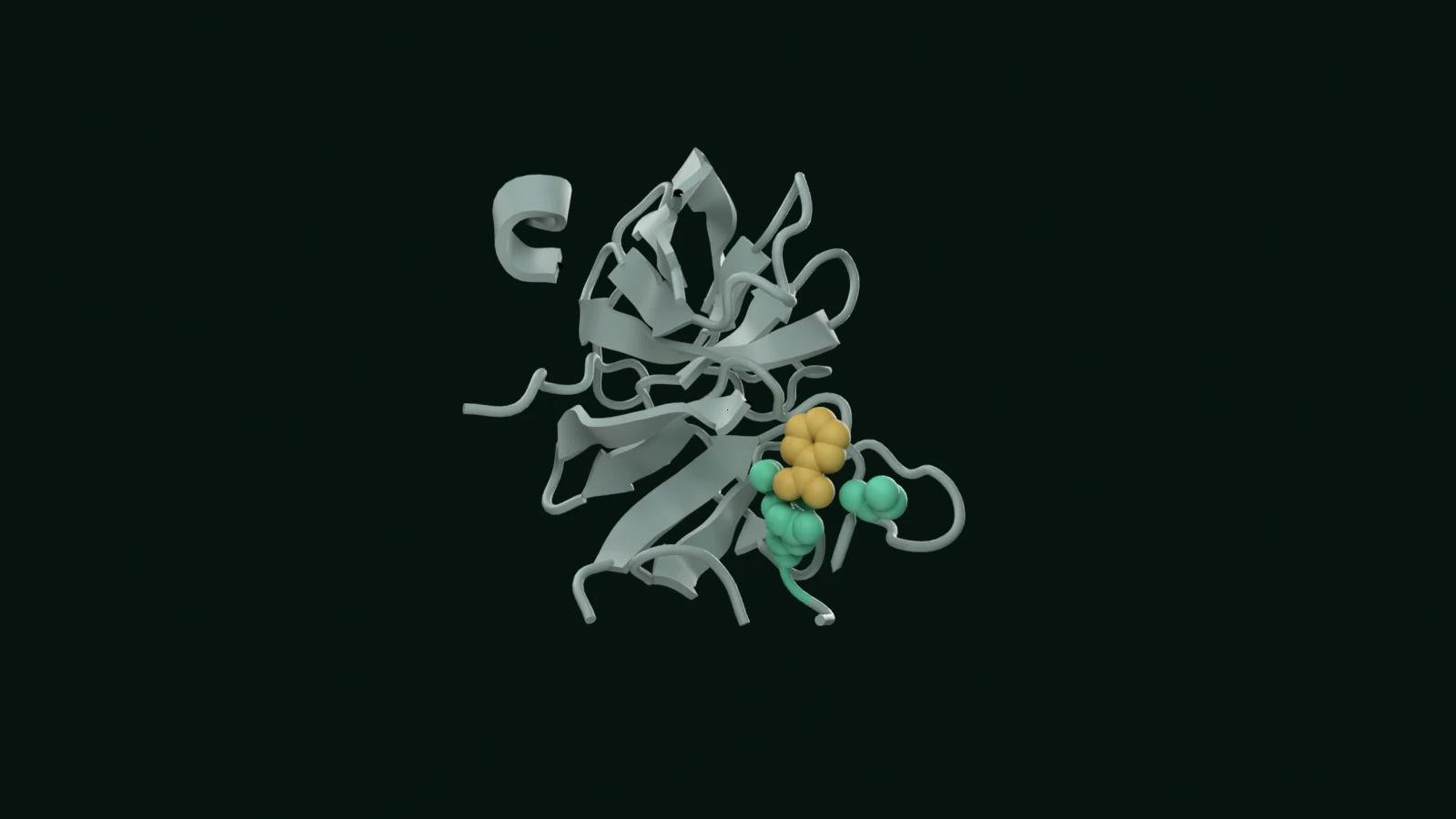
PDB 3PTB shows benzamidine bound to bovine trypsin in an X-ray structure at 1.70 Å. It provides a concrete pocket to inspect. The illustrative constants below do not come from those coordinates. Binding or displacement alone does not establish a biological effect.
Check your understanding Optional
A competitor halves ligand occupancy in the model. Has it necessarily halved the biological response?
At the bench
Move from defined binding relationships to measured concentration–response data, controls, curve fitting and uncertainty in Assay. Protein provides structural context for the target.
Open AssaySources and model notes
Structure-based views of resolved experimental coordinates. Cutaways, category colours, camera choices and guided highlights are authored for teaching. Coordinates remain fixed; highlights are not experimental trajectories or simulation outputs.
Experimental binding geometry; the numerical experiment is separate.
Equilibrium, free concentrations, saturation binding and competition assumptions.